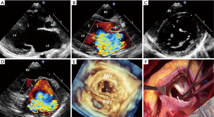
Figure 1.
MVC in a 14-year-old female. (A) 2D echocardiography in the parasternal long-axis view shows a break in the aortic mitral leaflet(arrows); (B) the parasternal long-axis view with color Doppler. The severe MR pass through the cleft and was directed posteriorly (arrows); (C) the parasternal short-axis view at the level of the mitral valve. A cleft in segment A2 of the anterior mitral leaflet (arrows); (D) the parasternal short-axis view at the level of the mitral valve. Color Doppler shows the severe MR coming through the cleft (arrows); (E) RT-3DE disclosed a cleft located in the midportion of the anterior mitral leaflet (arrows); (F) the intraoperative view from the left atrium onto the mitral valve. A cleft in segment A2 of the anterior mitral leaflet is shown (arrows). LA, left atrium; LV, left ventricle; RV, right ventricle; Ao, aortic; C, cleft; AMV, anterior mitral valve; PMV, posterior mitral valve.

