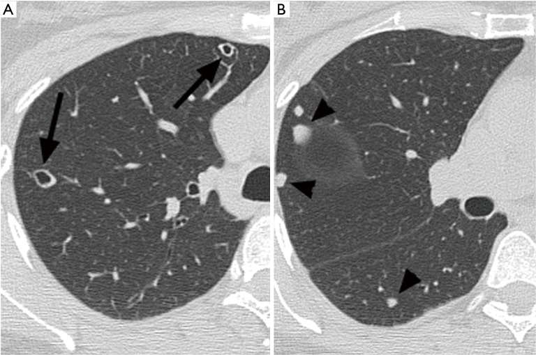
Figure 2.
Chest computed tomography (CT) at presentation. (A) Right lung on transverse thin section CT scan (1.0 mm collimation) obtained with lung window at the level of innominate vein shows a small thin walled cystic nodule (arrow) with well-defined solid nodule in in right upper lobe (arrowhead); (B) right lung on transverse thin section CT scan (1.0 mm collimation) obtained with lung window at the level of basal lobes demonstrates well demarcated several solid nodules with predominance of subpleural locations.

