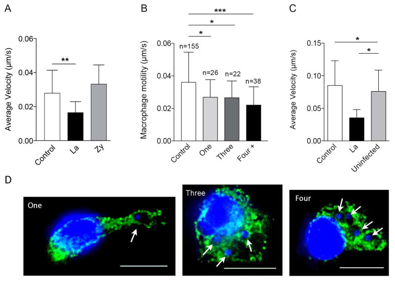
Figure 2. Inhibition of macrophage motility induced by L. amazonensis is not dependent on PV expansion, parasite load or secreted factors.
BMDM were exposed or not to L. amazonensis axenic amastigotes or to Zymosan particles for 1 h. BMDM were washed to remove extracellular parasites or particles, cultured at 34°C for another 24 or 48 h and assayed for their ability to migrate using phase-contrast time-lapse imaging. (A) Velocity of macrophages infected with L. amazonensis axenic amastigotes for 48 h carrying one, three, or more parasites. Data are representative of eight independent experiments. (B) Velocity of macrophages 48 h after internalization of L. amazonensis (La) or Zymosan particles (Zy). L. donovani (Ld), 48 h after infection. Data are representative of three independent experiments. (C) Effect of factors released into the medium by BMDM infected with L. amazonensis axenic amastigotes on macrophage motility 24 h after infection. Macrophage velocity was determined in non-infected macrophages in a separate culture (control) and in L. amazonensis-infected (La) and non-infected macrophages (uninfected) in the same culture. Data are representative of two independent experiments. (D) Images illustrating macrophages infected with one, three or four L. amazonensis axenic amastigotes for 48 h. Green, anti-Lamp1 antibodies; blue, DAPI DNA stain. Bars: 11 μm. * p < 0.05, ** p < 0.01, ** p < 0.001 (Student’s t test) compared with non-infected control BMDM.

