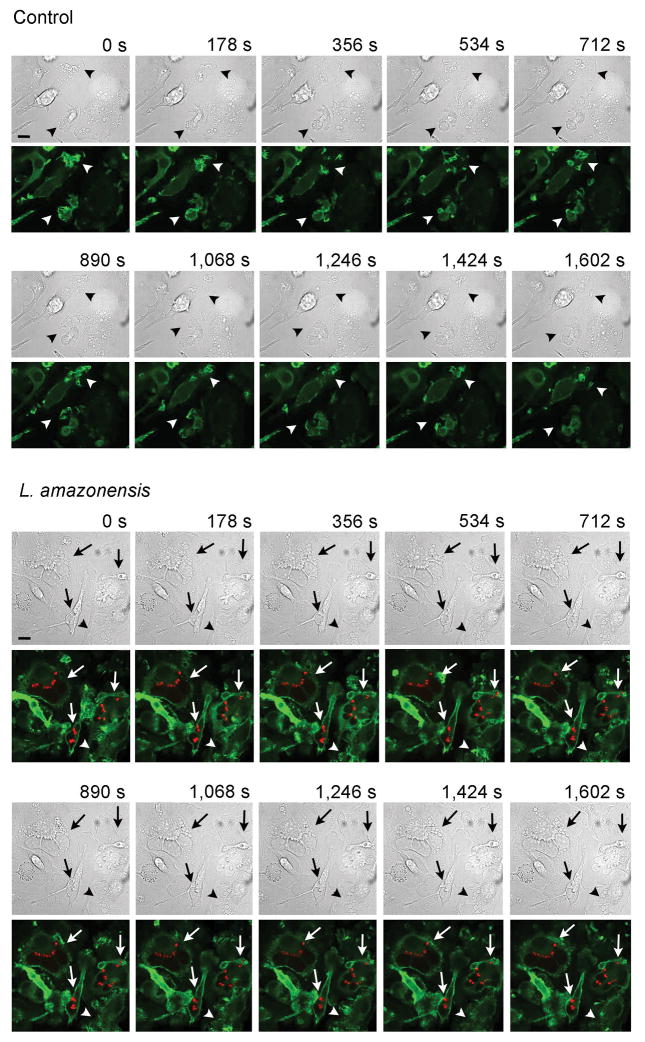
Figure 4. Differential actin filament dynamics during migration of macrophages infected with L. amazonensis.
BMDM from Lifeact mice were infected or not (control) with L. amazonensis axenic amastigotes for 1h. BMDM were then washed to remove extracellular parasites, cultured at 34°C for another 24 h, followed by fluorescence time-lapse image acquisition for 26.7 min. Arrowheads: F-actin (green) dynamics at ruffling leading edges of macrophages. Arrows: infected macrophages showing altered F-actin propagating waves at cell boundaries. Bars: 10 μm. See supplemental videos.

