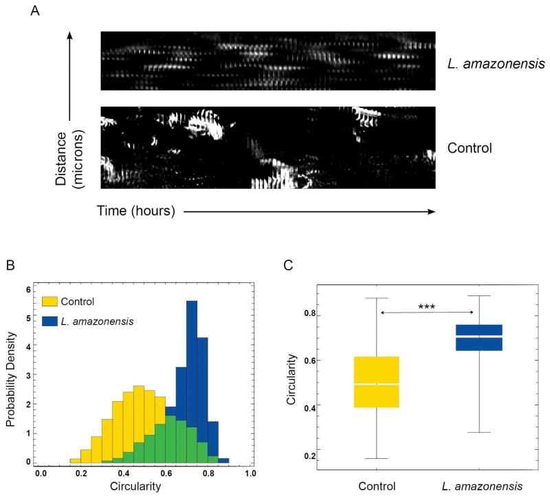
Figure 5. Actin filament dynamic properties are markedly altered in macrophages infected with L. amazonensis.
BMDM from Lifeact mice were infected or not with L. amazonensis axenic amastigotes for 1 h. BMDM were then washed to remove extracellular parasites, cultured at 34°C for another 24 h, followed by fluorescence time-lapse image acquisition. (A) Space/time plot of an actin propagating wave at the lamellipodia front over 200 frames indicating oscillating dotted actin polymerization waves in infected macrophages versus a propagating steady membrane wave front in an actively migrating control macrophage. The results shown are representative of four non-infected macrophages and five infected individual macrophages analyzed. (B) Shape dynamic analysis showing circular actin shape-like features in 25 infected macrophages versus line-like steady actin membranous features in 25 control macrophages. (C) Statistical analysis of the data in (B) indicating that infected macrophages show significantly more circular shape features compared to control macrophages. The boxes show the 50% confidence region from the median (black line). The bars cover a region with 99% confidence level from the median. *** p<0.01, n=25. The results shown are representative of two independent experiments.

