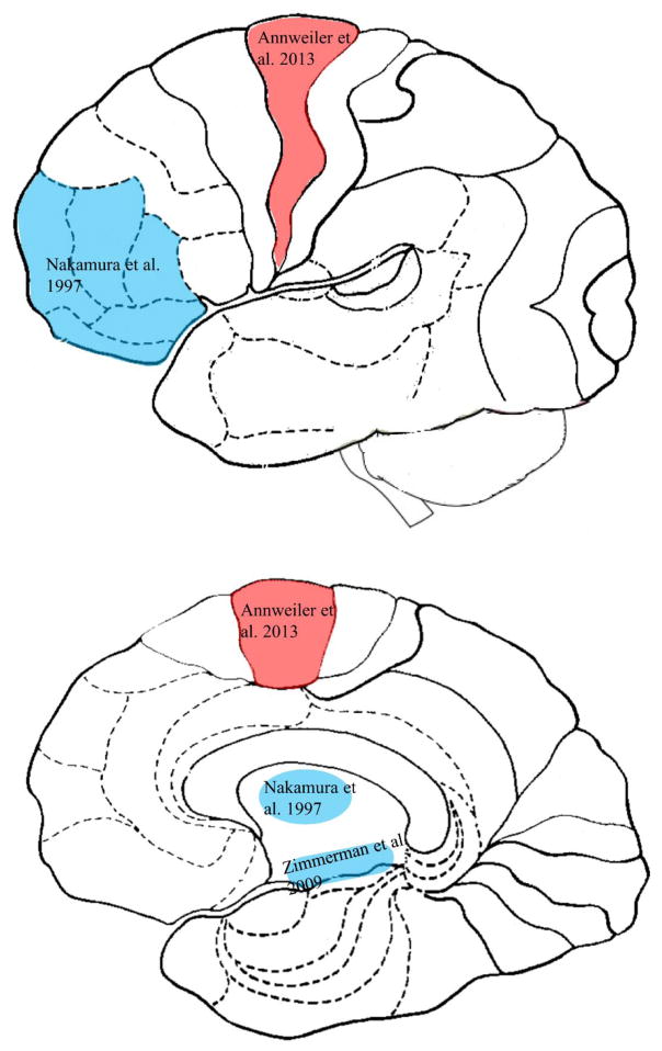
Figure 3. The brain map of gait variability in cognitive impairment or dementia.
Caption: Brain gray matter areas on sagittal view of the lateral cortex (top) and the medial cortex (bottom), that are associated with temporal gait variability (red) and spatial gait variability (blue) in older adults with cognitive impairment or dementia.

