Figure 1.
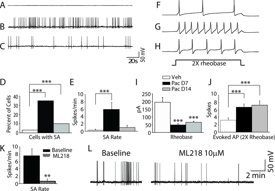
DRG neurons in rats with paclitaxel CIPN show hyper-excitability. A–C shows representative recordings of the spontaneous activity (SA) observed in (A) vehicle-, (B) day 7 paclitaxel-, and (C) day 14 paclitaxel-treated rats. The bar graphs in D and E shows the incidence and mean rate of SA in the paclitaxel groups was significantly higher than in the vehicle treated group. F–H shows representative action potential responses evoked at 2× rheobase in (F) vehicle-, (G) day 7 paclitaxel-, and (H) day 14 paclitaxel-treated rats. The bar graphs in I and J show that the mean current threshold at rheobase was significantly reduced in both paclitaxel treated groups and that the mean number of action potentials evoked at 2× rheobase was significantly higher. Figure K showed DRG neuron with spontaneous action potential (SA) isolated from segments at day 7 after paclitaxel treatment show frequency of SA was reduced by bath application of ML218 hydrochloride (10 µM). Figure L showed a representative recording traces before and after administration of ML218 hydrochloride (10 µM). **p < 0.01, ***p < 0.001.

