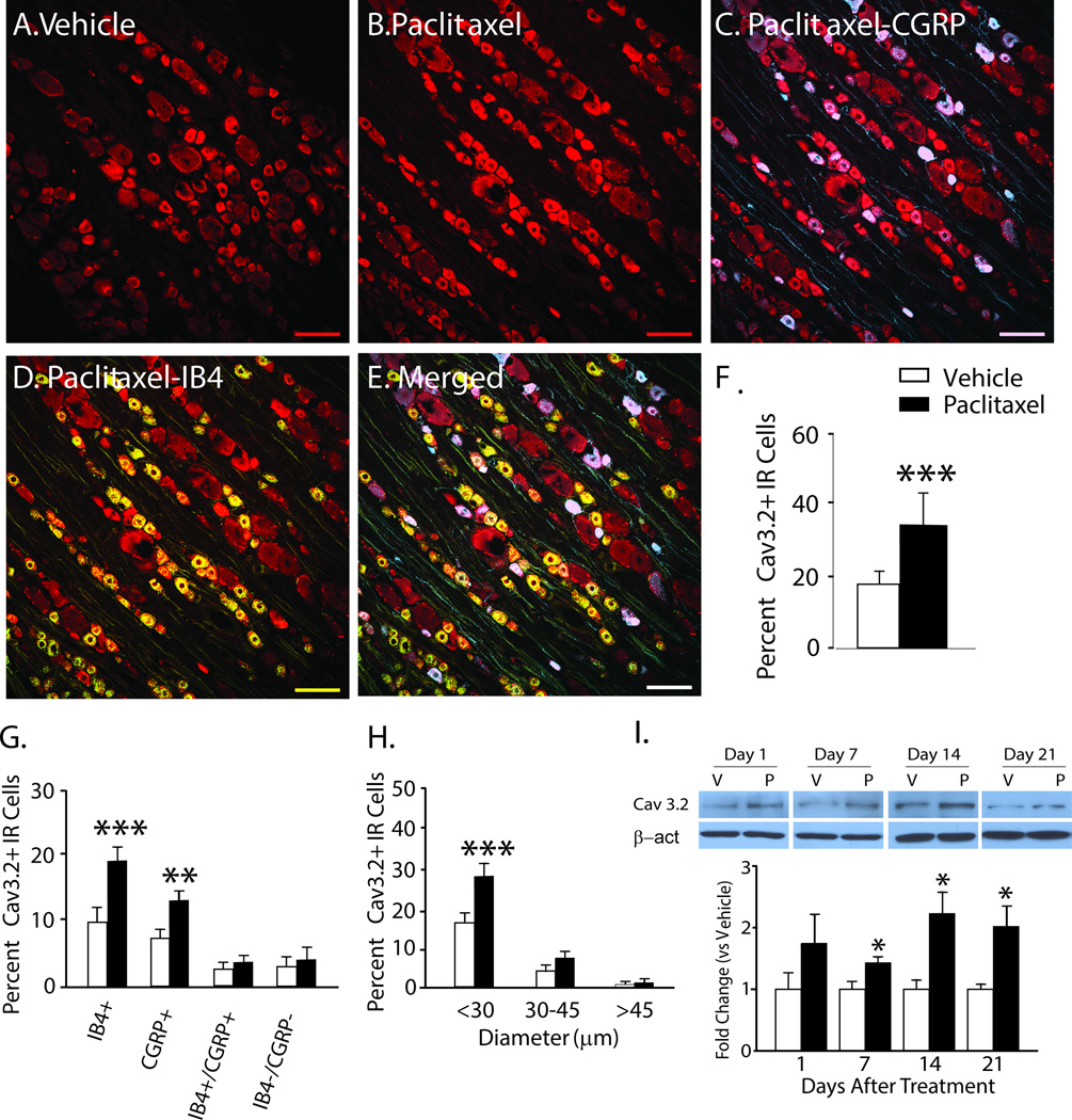
Figure 2.
The expression of Cav3.2 is increased in DRG in rats with paclitaxel CIPN. Representative Immunohistochemical (IHC) images are shown in A to E. The expression of Cav3.2 (red) in DRG is normally low in naïve (not shown) and vehicle-treated rats (A) becomes pronounced by day 7 after paclitaxel (B); and is co-localized in subsets of CGRP-positive (blue) neurons (C, co-localization indicated in purple) and IB4-positive (green) neurons (D, co-localization indicated in yellow), as well as in a large percentage of neurons negative for both IB4 and CGRP (E, overall merged). The bar graphs in F to H summarize the grouped IHC data and show that the overall number of Cav3.2 positive neurons (F) was significantly higher in the paclitaxel-treated rats (black bars) than in the vehicle-treated rats (white bars); and these increases were significantly higher in IB4 positive and CGRP positive neurons (G) and in neurons with diameters less than 30 µm (H). In I representative western blot images and bar graph grouped data confirm that the expression of Cav3.2 was significantly increased in the DRG at day 7 through day 21 after chemotherapy. n = 4/group in both the IHC and western blot experiments. Scale bar = 100µm. β-act= beta-actin; V = vehicle; P = paclitaxel. *p < 0.05; **p < 0.01; ***p < 0.001.

