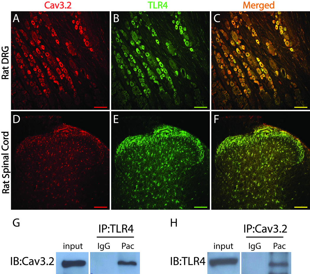
Figure 4.
Cav3.2 is co-localized with TLR4 in rat DRG and spinal cord. A–F, Representative Immunohistochemistry (IHC) images showing Cav3.2 (red) is co-localized (yellow) with TLR4 (green) in rat DRG neurons (A-C) and rat spinal cord cells (D–F). G and H, The total protein extracted from rat DRG at the day 7 paclitaxel was used for immunoprecipitation (IP) with an anti-TLR4 antibody and immunoblotting (IB) with an anti-Cav3.2 antibody (G) or immunoprecipitation with an anti-Cav3.2 antibody and immunoblotting with an anti-TLR4 antibody (H). IgG was used to confirm the specificity of the antibodies. Scale bar, 100 µm.

