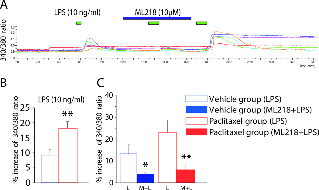
Figure 6.
T-type calcium channels are activated by LPS via TLR4 in DRG neurons. A, Representative calcium imaging results showing the change in 340/380 ratio in dissociated DRG neurons following perfusion of LPS alone (green bar), followed by LPS plus ML218 hydrochloride (blue bar) and then LPS alone again. Each colored line represents a single neuron, and the time of each application is indicated by the bars above the traces. B and C, the bar graphs show the grouped results of experiments performed to determine the effects of LPS on DRG neurons alone in the vehicle and paclitaxel groups (B) and the effects of ML218 hydrochloride on LPS-induced intracellular calcium enhancement (C). Treatment with ML218 hydrochloride significantly reduced calcium entry stimulated by LPS. *p < 0.05; **p < 0.01.

