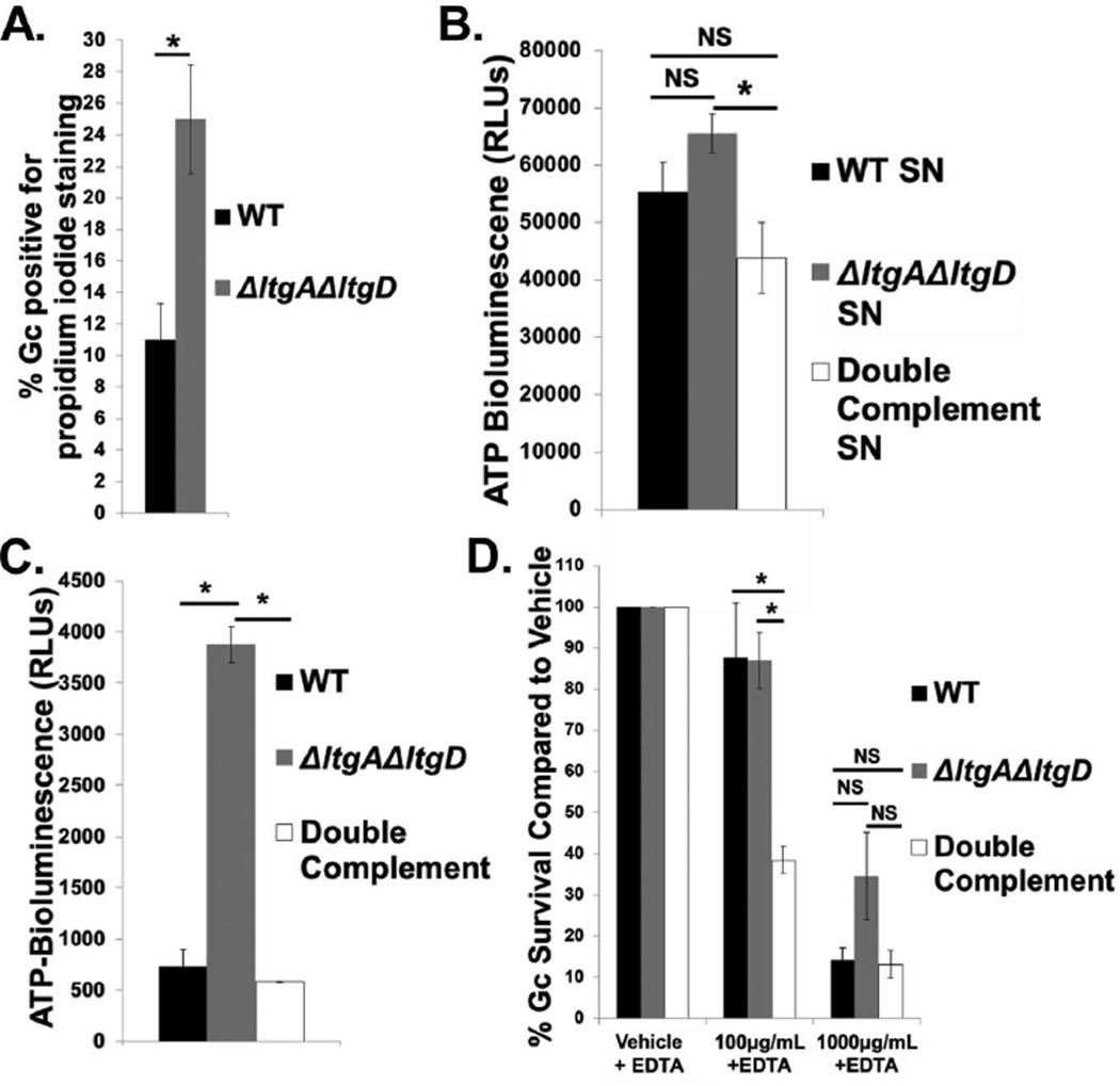
Figure 5. LtgA and LtgD contribute to Gc membrane integrity.
A. WT and ΔltgAΔltgD double mutant Gc were treated with propidium iodide to label membrane-permeant Gc and Syto9 to label total Gc. Percent Gc positive for propidium iodide staining was calculated by dividing total propidium iodide positive Gc by total Gc. n = 5 biological replicates.
B. Supernatant (SN) was harvested from WT, ΔltgAΔltgD double mutant, and ltgA+ltgD+ double complement Gc at an equivalent optical density and incubated with luciferin and luciferase from an ATP determination kit. Bioluminescence was measured with a luminometer as relative light units (RLUs). n = 3 biological replicates.
C. WT, ΔltgAΔltgD double mutant, and ltgA+ltgD+ double complement Gc at an equivalent optical density were incubated with Toxilight reagent containing ADP, luciferin, and luciferase for 5 min at room temperature. Positive extracellular adenylate kinase activity produced bioluminescence, reported in RLUs. n = 3 biological replicates.
D. WT, ΔltgAΔltgD double mutant, and ltgA+ltgD+ double complement Gc were exposed to increasing concentrations of human lysozyme or vehicle control in the presence of 1mM EDTA for all conditions, for 30 min. Gc survival is expressed as in Fig. 3A. n = 6 to 9 biological replicates.
In all cases, *P<0.05; two-tailed t-test.

