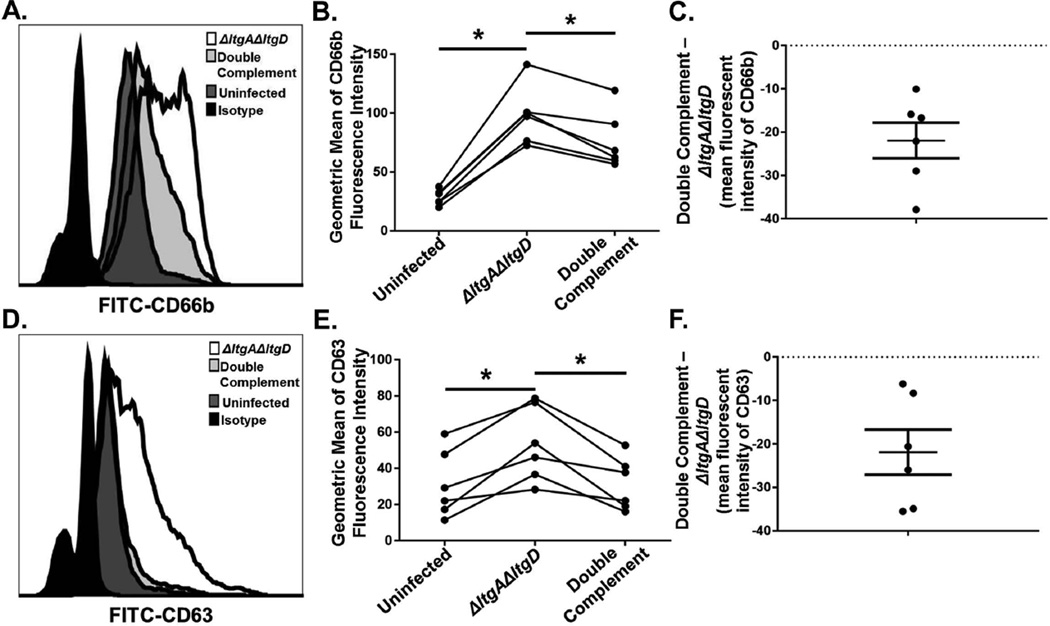
Figure 7. Increased secondary and primary granule exocytosis in primary human neutrophils exposed to ΔltgAΔltgD double mutant Gc.
IL-8-treated, adherent human neutrophils were left untreated, exposed to ΔltgAΔltgD double mutant, or exposed to ltgA+ltgD+ double complement Gc for 1 hr. Neutrophils were subsequently stained with FITC-CD66b as a marker for secondary granule exocytosis (A) or FITC-CD63 as a marker for primary granule exocytosis (D) and analyzed by flow cytometry. The geometric mean fluorescence intensity for CD66b (B) and CD63 (E) was calculated from the granulocyte population. The differences of mean fluorescence intensity of ΔltgAΔltgD mutant Gc from double complement Gc were determined for CD66b (C) and CD63 (F); data are shown as mean ± SEM. *P < 0.05; paired, two-tailed t-test, n = 6 independent experiments.

