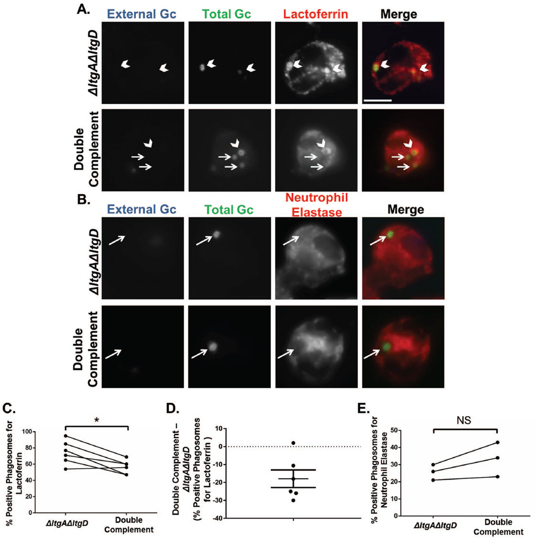
Figure 8. Increased delivery of secondary granules to neutrophil phagosomes containing ΔltgAΔltgD double mutant Gc.
Neutrophils were exposed to CFSE-labelled ΔltgAΔltgD double mutant or ltgA+ltgD+ double complement Gc (green) for 1 hr. Extracellular bacteria were labelled with an anti-Gc antibody (blue). Phagosomes were labelled with an anti-lactoferrin antibody to identify secondary granule-positive phagosomes (A) or an anti-neutrophil elastase antibody to identify primary granule-positive phagosomes (B). Phagosomes were considered positive when greater than 50% of the ring around the bacterium is positively stained for the antibody. Scale bar, 5 µm. Percent phagosomes positive for lactoferrin (C) and neutrophil elastase (E) were quantified by dividing positive phagosomes by total phagosomes. D depicts the difference in lactoferrin-positive phagosomes between ΔltgAΔltgD double mutant and double complement Gc as mean ± SEM. *P<0.05 for ΔltgAΔltgD compared to double complement; two-tailed t-test, n= 3 to 6 independent experiments.

