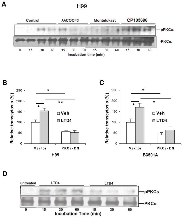
Figure 6. cPLA2α and cysteinyl LTs contribute to C. neoformans traversal of the HBMEC monolayer via PKCα.
(A) HBMEC incubated with C. neoformans strain H99 at 37°C for various times in the presence of inhibitors/antagonists or vehicle control were immunoprecipitated with PKCα antibody, and then assessed for phospho-PKC by Western blotting with phospho-PKC antibody. The HBMEC lysates were examined for the total amounts of PKC.
(B and C) Effect of LTD4 on penetration of C. neoformans strain H99 (B) or B-3501A (C) across monolayer of HBMEC transfected with dominant-negative PKCα construct. Subconfluent HBMEC in transwell insert were infected with adenovirus Ad5CA dominant negative-PKC-α (DN) or vector control (Vec) at MOI of 50. HBMEC were pretreated with 1 μM LTD4 for 30 min followed by transcytosis assays. 0.5% ethanol was used as vehicle control (Veh). Data shown are mean ± SEM. Each experiment was performed in triplicate. *P <0.05; **P <0.01, Student’s t test.
(D) LTD4 is involved in PKCα activation in HBMEC. HBMEC incubated with LTD4 (1 μM) or LTB4 (1 μM) at 37°C for various times (min) were immunoprecipitated with PKCα antibody, and then assessed for phospho-PKC by Western blotting with phospho-PKC antibody. The HBMEC lysates were examined for the total amounts of PKC.

