
Figure 3.
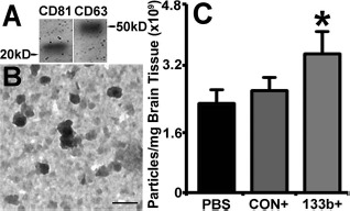
MicroRNA 133b (miR-133b)-enriched exosomes increased exosomes in brain ischemic boundary zone (IBZ). Western blot shows that the common exosomal proteins CD63 and CD81 are present in the exosome extraction from the IBZ (A). The transmission electron microscopic (TEM) images show the morphology of exosomes presented in the brain extracellular (B), within a size range of 30–100 nm. qNano quantification data show that compared to the multipotent mesenchymal stromal cells (MSCs) infected with blank vector (Ex-Con) treatment and phosphate-buffered saline (PBS) control, the extracellular exosomes present in the IBZ significantly increased after miR-133b-overexpressing MSC (Ex-miR-133b+) treatment (C). PBS, middle cerebral artery occlusion (MCAO) rats treated with PBS; CON+, MCAO rats treated with Ex-Con; 133b+, MCAO rats treated with Ex-miR-133b+. ∗p <0.05, compared to Ex-Con. Mean ± standard error (SE), n = 6/group. Scale bar: 100 nm.