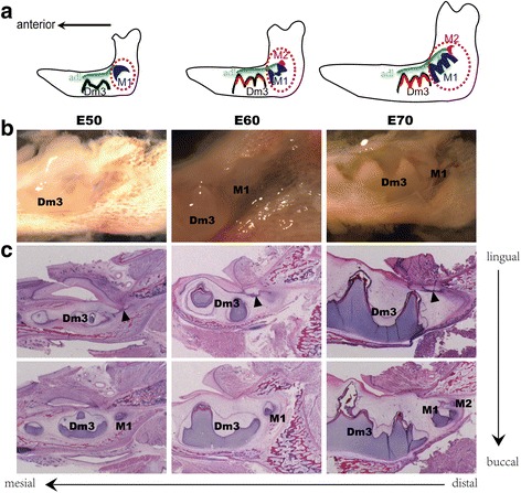
Fig. 1.

Sequential initiation pattern of mandibular additional molar during morphogenesis in the miniature pig. a Schematic representation of additional molar germs (dashed lines) in mandible dissected from E50, E60 and E70 miniature pig embryos for transcriptome analysis, showing a sequential initiation pattern. b Macro view of additional molar germ in miniature pig mandible at the different stages. c Serial sagittal histological sections (haematoxylin and eosin staining) show the morphogenesis of additional molars at the different stages and additional dental lamina (arrowhead) from the distal extension of the primary dental lamina. adl, additional dental lamina. Dm3, the third deciduous molar. M1, the first additional molar. M2, the second additional molar
