Fig. 8.

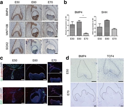
Gene expression in SHH, WNT and TGF-β pathways during cascade initiation of additional molars. a Immunohistochemistry of the expression of genes related to TGF-β, WNT, SHH and MAPK pathways. BMP4 expression (top panel) mainly resides in the inner enamel epithelium and outer enamel epithelium at E50; is located in the inner enamel epithelium, outer enamel epithelium, dental papilla and additional dental lamina at E60; and then is concentrated in the inner enamel epithelium at E70. WNT10b shows a similar expression pattern with BMP4 (middle panel). SOX2 expression (bottom panel), located in the inner enamel epithelium and outer enamel epithelium at E50, mainly occurs in additional dental lamina at E60 and is visible in inner enamel epithelium and additional dental lamina at E70. SOX2 expression in additional dental lamina shows an asymmetric distribution. Positive expression is visualized by DAB (brown) and counterstained with haematoxylin (blue). Lingual is to the left. Scale bars, 200 μm. b RT-qPCR of the expression of BMP4 and SHH showing dynamic changes consistent with immunohistochemistry results. Error bars indicate s.d. three biological replicates. c Immunofluorescence shows the relationship among the SHH, WNT and TGF-β pathways. Immunofluorescence of Gli1 (green) and β-catenin (red) shows their co-expression in inner enamel epithelium, outer enamel epithelium and additional dental lamina (top panel); co-immunostaining of SHH (green) and OSR2 (red) in inner enamel epithelium, outer enamel epithelium, dental papilla and additional dental lamina. Lingual is to the right. Scale bars, 500 μm. d mRNA expression of BMP4 and an important WNT pathway transcription factor TCF4 detected by in situ hybridization at E50 and E70 of M1 (blue). At E50, BMP4 is expressed in the inner enamel epithelium especially in the enamel knot and in the mesenchyme cells close to the epithelium. At E70, BMP4 is mainly expressed in the inner enamel epithelium, similar to immunofluorescence results. TCF4 is expressed in the inner enamel epithelium, outer enamel epithelium and dental papilla at E50 and only in dental papilla at E70. Black dashed lines mark the boundary of the tooth bud epithelium. Scale bars, 100 μm
