Fig. 7.

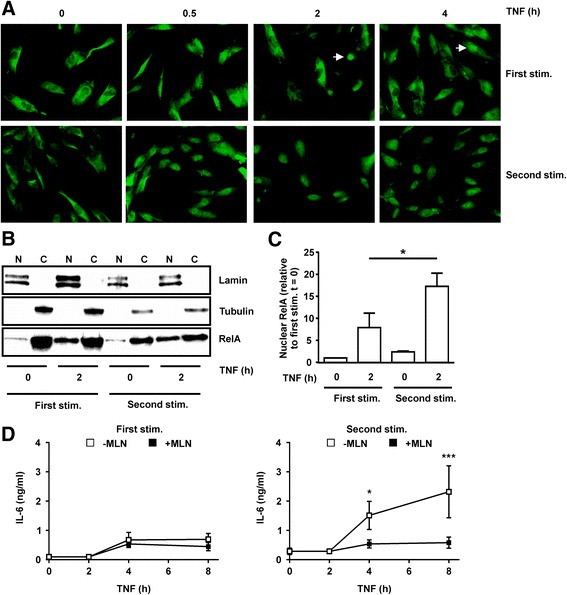
Prolonged nuclear factor (NF)-κB activation in re-stimulated BJ cells contributes to enhanced IL-6 biosynthesis. a BJ fibroblasts were seeded on chamber slides and stimulated with TNFα (10 ng/mL) for varying times, with or without prior exposure to TNFα for 24 h and rest in the absence of TNFα for 24 h. RelA was detected by immunofluourescence. Images are representative of two independent experiments. White arrows indicate cells in which RelA is principally nuclear. b BJ fibroblasts were untreated or stimulated (stim) with TNFα (10 ng/mL) for 2 h, with or without priming as above. Nuclear (N) and cytoplasmic (C) fractions were prepared and RelA was detected by western blotting. Lamin A/C and tubulin were blotted in order to validate the subcellular fractions. Results are representative of four independent experiments. c Nuclear RelA was quantified, normalised against the Lamin A/C loading control, and plotted relative to the level at t = 0 in the first TNFα challenge. Mean ± SEM, n = 4; *p < 0.05 (Mann-Whitney U test). d BJ fibroblasts were stimulated (left) or re-stimulated (right) with 10 ng/mL of TNFα, and 100 nM MLN4924 or vehicle control (0.1% dimethyl sulfoxide (DMSO)) was added after 2 h. Supernatants were collected at the time points indicated, and IL-6 measured by ELISA; n = 6; *p < 0.05, ***p < 0.005; Student t test
