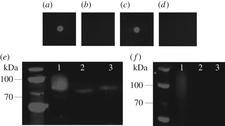
Figure 5.
Glycosylation analysis of AtaC1866–2428 alongside NGT and α6GlcT by anti-dextran and anti-HIS western blots. Top panel: anti-dextran dot blot; (a) dextran; (b) AtaC with NGT K441A and functional α6GlcT; (c) AtaC with functional NGT and α6GlcT; (d) AtaC with functional NGT only. Bottom panel: anti-HIS and anti-dextran Western blots (e,f, respectively); lane 1, AtaC with functional NGT and α6GlcT; lane 2, AtaC with NGT K441A and functional α6GlcT; lane 3, AtaC with functional NGT only.

