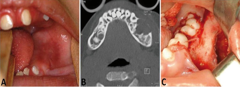
Figure 1.
A) Intraoral view showing a buccal expansion affecting the left mandibular region. B) CT (axial section, bone window) displaying a hypodense image with bucco-lingual bony expansion and cortical bone thinning. C) Intraoperative image of the lesion showing impacted teeth and the erosion of the cortical bone.

