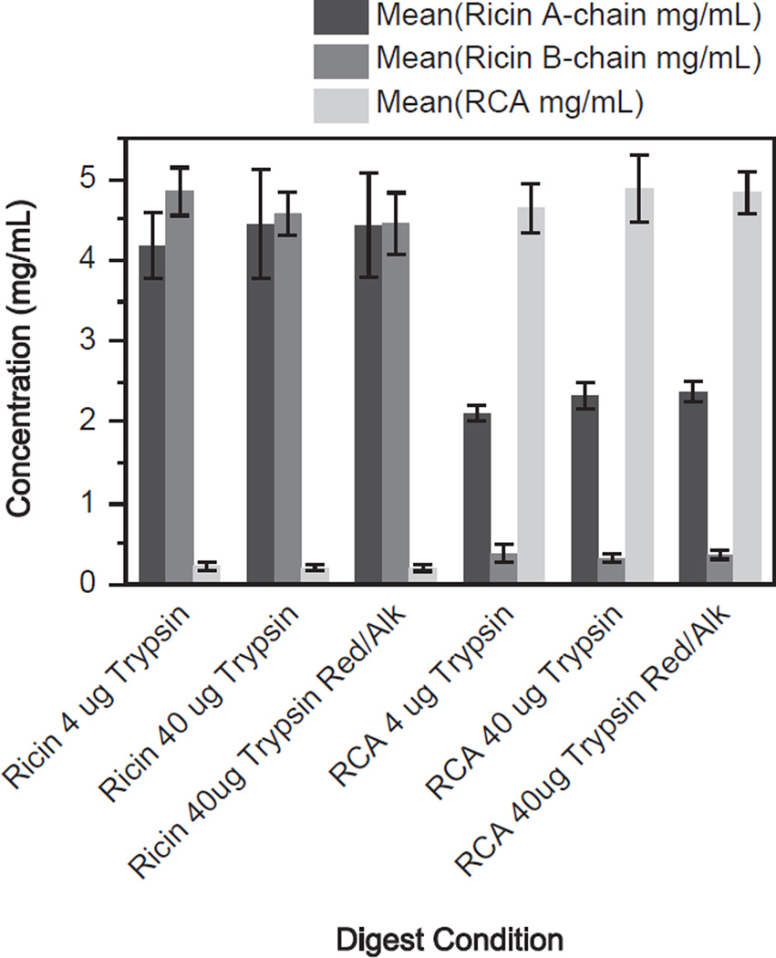
Fig. 1.
Measurement of purified ricin and RCA standard concentrations by isotope dilution mass spectrometry. Three different tryptic digestion conditions using 4 µg, 40 µg and 40 µg with reduction and alkylation were used to digest ricin and RCA. Quantity of protein was determined using a peptide based standard calibration curve. The ricin A-chain peptides are denoted in dark gray, ricin B-chain peptide is medium gray and the RCA A-chain peptides are denoted in light gray. Error bars show one standard deviation from the mean for three replicates.

