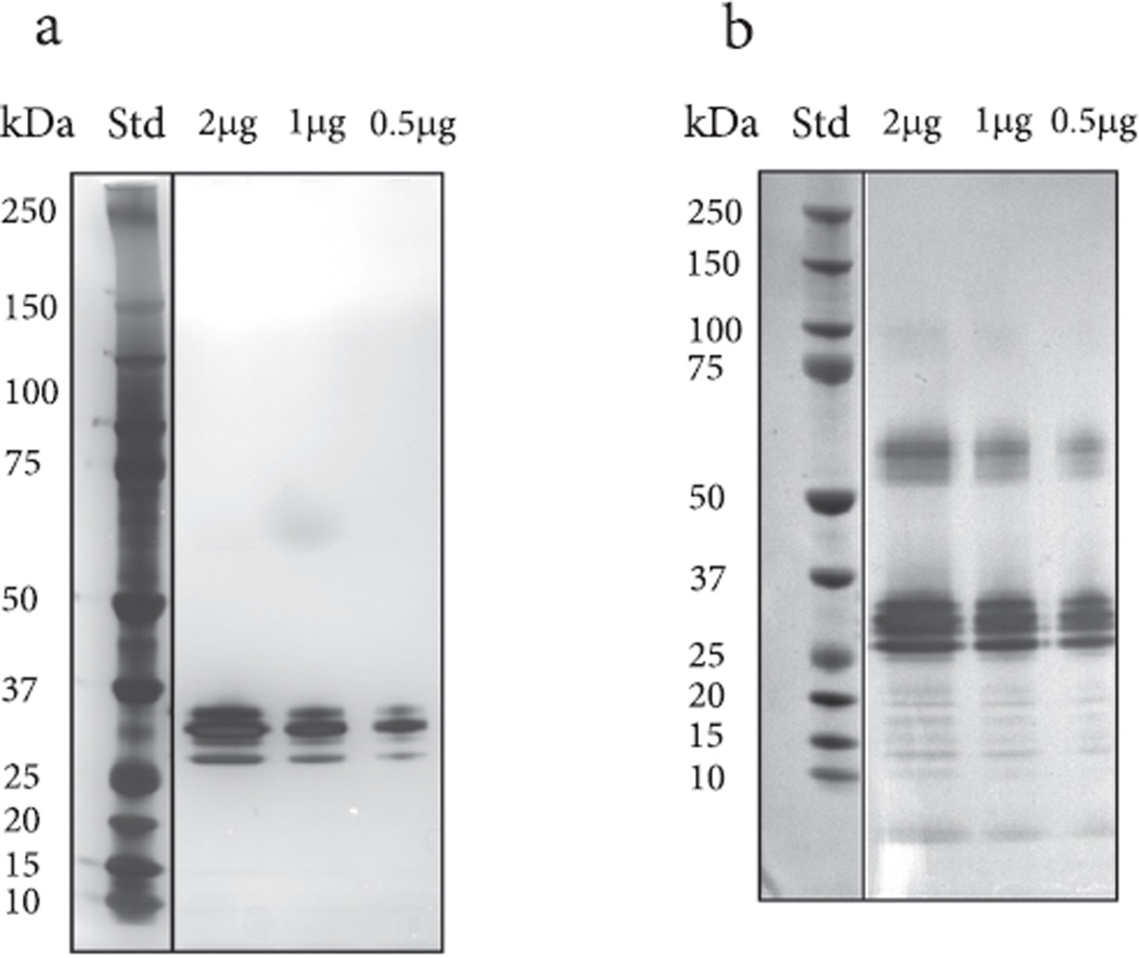
Fig. 7.
a. Separation and visualization of the ricin reference standard by one dimensional SDS-PAGE and silver staining. Molecular weight markers are in the left lane and 2, 1 and 0.5µg of the reference ricin are in the subsequent lanes. Individual ricin chains are likely located between 25 and 37 kDa b. is a duplicate gel use in the Western blot analysis. A series of immuno-reactive bands to the goat anti-ricinus communis antibody are seen between 50 and 75 kDa, 25–37 kDa and 10–20 kDa.

