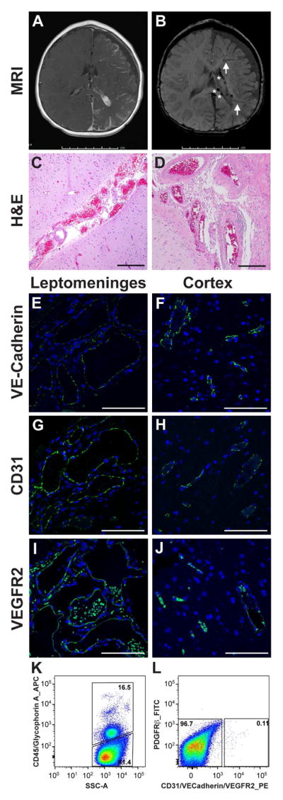
Figure 1. Brain specimens from two SWS patients.

(A) Post-contrast axial T1-weighted MRI in patient 1 shows extensive left hemisphere leptomeningeal enhancement, left choroid plexus hypertrophy and marked left cerebral atrophy. (B) Axial susceptibility-weighted MRI in patient 1 shows enlarged medullary veins (arrows) and dilated deep veins (*). (C) Hematoxylin and eosin (H&E) staining of patient 1 brain tissue section shows abnormal clusters of leptomeningeal vascular channels of variable calibers. The underlying cortex demonstrates focal loss of neurons in layer 2 and subpial gliosis. Scale bar, 100μm. (D) H&E staining, viewed at higher magnification, shows some of these vascular walls are thick and rigid. Scale bar, 200μm. The histologic findings are consistent with leptomeningeal angiomatosis. (E–J) Anti human VE-cadherin, anti-human CD31 and anti-human VEGFR2 staining highlights enlarged vessels in leptomeningeal angiomas ( E,G and I, green) and shows positive staining predominately in the endothelium in the cortex (F,H and J, green) in patient 1. Nuclei counterstained with DAPI (blue). Scale bar, 100μm. (G–H) Fluorescence-activated cell sorting of freshly isolated brain cells from patient 2. Cells were sorted into hematopoietic and non-hematopoietic cells based on CD45/Glycophorin A expression (G). Non-hematopoietic cells were further fractioned into endothelial cells (using a set of anti-endothelial antibodies against CD31, VE-Cadherin and VEGFR2), PDGFRβ-positive cells and cells negative for all markers (H). Percentages of cells in each population are shown.
