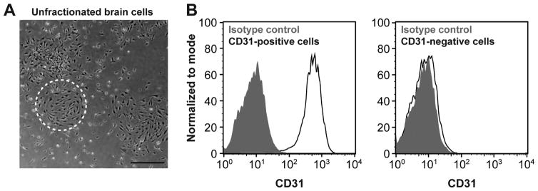
Figure 2. Cells isolated from SWS brain lesion.
(A) Primary culture from patient 1 brain lesion. Dashed circle highlights cells with endothelial morphology. Scale bar, 50μm (B) Flow cytometric analysis of cells in the primary culture after selection using anti-CD31-coated magnetic beads: CD31-positive cells and CD31-negative cells. Black lines depict cells incubated with anti-CD31 antibody; shaded grey areas are cells incubated with isotype-matched control antibody.

