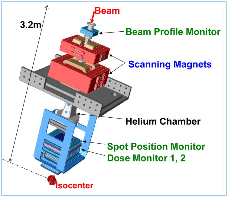
Figure 7.
A Scanning beam nozzle (Hitachi at MDACC). The thin beamlets of a sequence of energies entering the nozzle are spread laterally by a pair of x- and y-magnets to create a three-dimensional pattern of dose distribution. Magnet strengths are adjusted to confine the Bragg peaks of beamlets (“spots”) to within the target volume. Intensities of beamlets, computed using a treatment planning system, are optimized in order to conform the high and uniform dose pattern to the target volume and appropriately spare critical normal tissues. Various monitoring systems ensure that the characteristics of the proton beam are within specifications and that the requisite dose is accurately delivered. Part of the path from the beamlet entry position to the isocenter is replaced with a helium chamber to reduce lateral dispersion of the scanning beamlet in air.

