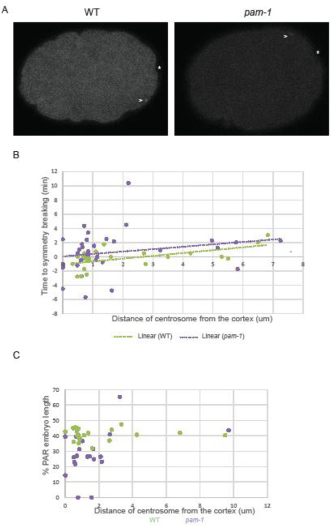
Figure 3. The timing of centrosome-cortical contact does not correlate with polarity defects.
A) In pam-1 mutants, the centrosome (γ-tub::GFP) contacts the cortex earlier in the cell cycle than WT. The times were measured back from nuclear envelope breakdown (NEBD). Images show the start of centrosome-cortical contact and a pam-1 mutant embryo at a time comparable to the start time for WT. Posterior is to the right in all images. B) Both the start and end of centrosome-cortical contact are significantly different. (WT n=39; pam-1 n=43) Standard deviations are shown. * p≤0.01; *** p≤0.0001. C) End of centrosome (arrowhead) contact times in wild-type occurred during the pronuclear phase as viewed by histone::GFP. In pam-1 mutants, end of centrosome contact usually occurred prior to the appearance of decondensed pronuclei. D) pam-1 mutant embryos were separated by extent of PC and end of centrosome contact was compared (no PC n=17; weak PC n=22; PC n=4). The mean in all three classes were different from WT. * p≤0.01; *** p≤0.0001. Box plots show range of data with the box representing the upper and lower quartile and the median. Whiskers show the max and min.

