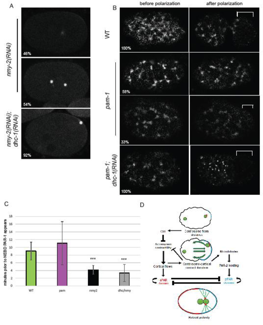
Figure 5. Centrosome-cortical contact enhances polarity through both the actomyosin-dependent and microtubule-dependent pathways.
A) In embryos with no cortical flows, in the absence of NMY-2, by NEBD PAR-1 localizes to a small cortical patch 46% of the time. PAR-1::GFP and γ-tub::GFP. When dhc-1 is also inactivated, 92% of embryos now have PAR-1 localization at NEBD. B) pam-1 embryos have sparse NMY-2 foci in the cortex as compared to WT. While WT embryos show clearing of NMY-2 foci from the posterior after polarization, 58% of pam-1 mutants show no clearing and 33% show weak clearing. When dhc-1 is inactivated in pam-1 mutants, all embryos now show NMY-2 clearing from the posterior. NMY-2::GFP clearings shown with a bracket; posterior is to the right. C) PAR-1::GFP localization to the posterior occurs later when flows are absent. * p≤0.05; *** p≤0.0001 Standard deviation is shown. (WT: n=32; pam-1: n=44; nmy-2(RNAi): n=8; nmy-2, dhc-1(RNAi): n=10) D) A revision to the model of Motegi and Seydoux (2013). Centrosome-cortical contact enhances polarization through the actomyosin-dependent and the microtubule-dependent pathways. This contact is necessary when flows are weak or absent, but is redundant when cortical flows are strong.

