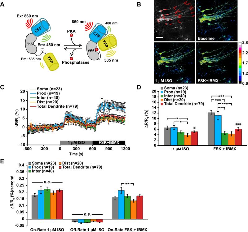
Figure 2. PKA activity in CA1 neurons is greater in somata and proximal dendrites during β-adrenergic receptor activation.
(A) Left: Cartoon of how AKAR3 functions as a substrate and reporter of PKA activity. Right: Representative example of PKA activity in CA1 neurons of brain slices. (B, C and D) (B) Average timecourse, (C) average peak amplitude of PKA activity during application of 1 μM isoproterenol (F(3,98) = 3.837, p = 0.0121) ), (#p = 0.0491) or FSK + IBMX (F(3,98) = 5.373, p < 0.0001), (###p < 0.0001) and (D) average rates of PKA activity during application of 1 μM isoproterenol or FSK + IBMX (F(3,98) = 4.371, p = 0.0062). Data are mean ± SEM. Neuronal compartments were analyzed by one-way ANOVA and Tukey's Test (*p < 0.05, **p < 0.01, ***p < 0.001). Soma vs total dendrites were analyzed by unpaired, two-tailed t-test with p-value as indicated. Image scale bars are 40 μm.

