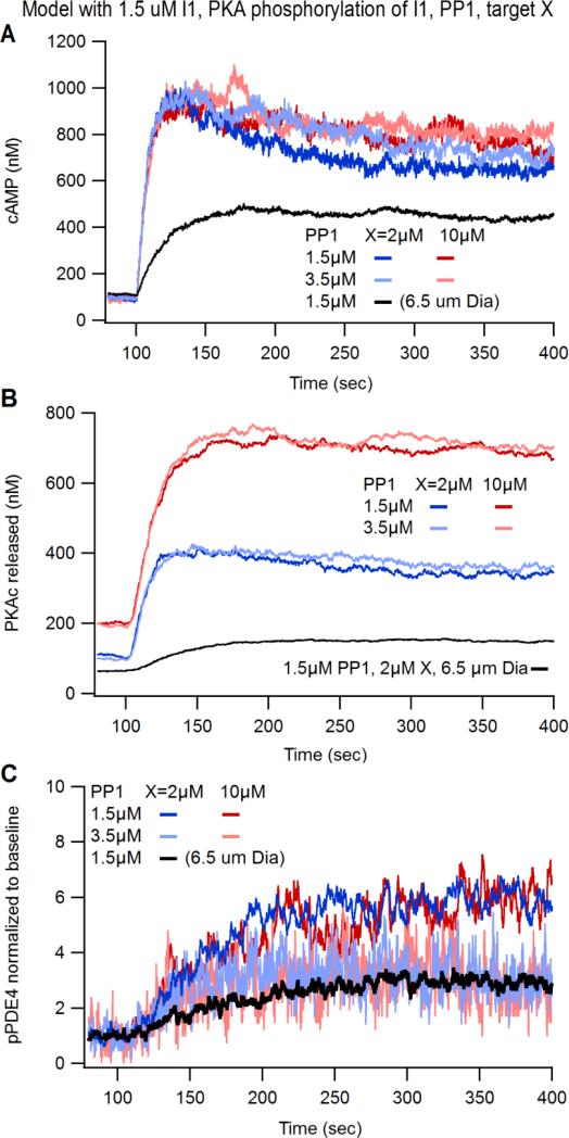
Figure 5. Computational modeling determines that protein phosphatase 1 is a potential mechanism suppressing PKA activity in small dendritic compartments and excess PKA substrate does not affect PKA signaling dynamics.
(A-C) The computational model defined in Fig. 3A was modified to include different concentrations of protein phosphatase 1 (PP1) and various concentrations of PKA substrate (molecule “X”). (A) cAMP transients were not affected by different concentrations of PP1 or X. (B) PKAc Released increases with greater concentrations of X but were unaffected by greater PP1. (C) The normalized phosphorylation status of PDE4 was not affected by different concentrations of X, but was reduced with greater concentrations of PP1.

