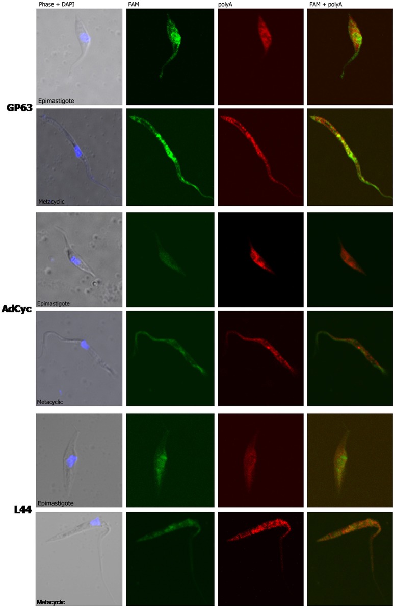
Figure 9.
mRNA localization in epimastigote and metacyclic trypomastigotes of T. cruzi. Confocal images of FISH assays of representative parasites using probes for: a surface protease GP63 transcript, ID: TcCLB.511211.90, GP63, upper panel. A receptor-type adenylate cyclase transcript, ID: TcCLB.511043.60, AdCyc, middle panel; and the 60S ribosomal protein L44 transcript, ID: TcCLB.507105.40, L44 bottom. At least 30 parasites were analyzed for each probe in each condition. Staining of epimastigotes (upper panels) and metacyclic trypomastigotes (bottom panels) are presented. These probes (See Table S2) were 5′ labeled with 6-FAM (green). For the detection of polyA tailed transcripts a 5′ Cy5 labeled polyT probe was used (red). DNA was visualized by DAPI staining (blue). Phase contrast images were obtained. Magnification 120x.

