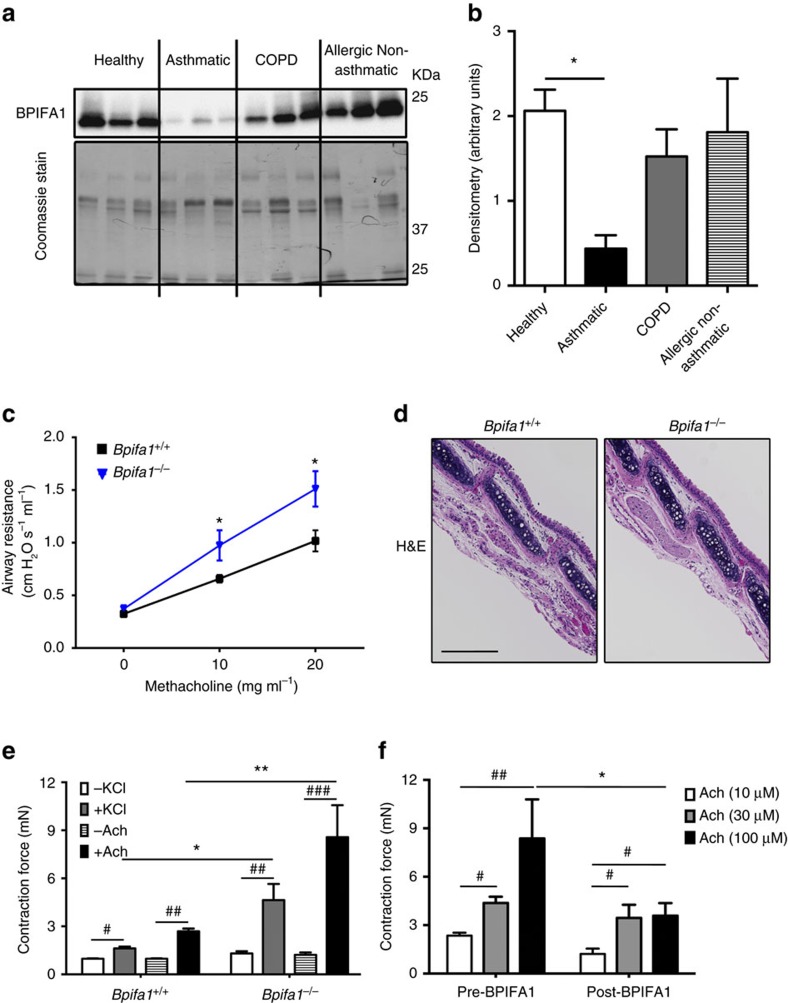
Figure 1. BPIFA1 is diminished in asthmatic airways and is associated with airway hyperresponsiveness (AHR) in mice.
Induced sputum was collected from healthy normal controls, asthmatic and chronic obstructive pulmonary disease (COPD) patients, and allergic non-asthmatics. (a) Representative immunoblots of BPIFA1 (upper) and coomassie loading control (lower). (b) Mean densitometry of BPIFA1 normalized to total protein. (n=6 subjects per group). (c) Evaluation of peripheral airway resistance by flexiVent after methacholine challenge in Bpifa1−/− and Bpifa1+/+ littermate controls. (d) Representative haematoxylin and eosin (H&E) staining of tracheas from Bpifa1−/− and Bpifa1+/+ mice (n=3 mice/genotype). Scale bar is 200 μm. (e) Tracheal rings (n=6 mice per genotype) were extracted from Bpifa1+/+ and Bpifa1−/− mice and mounted onto wire myographs. Contraction was measured under both resting and agonist (KCl and Ach)-induced conditions. (f) Contractile force was measured pre- and post-bilateral BPIFA1 addition to the bath 1 h before agonist addition (all n=6). Data in b,c,e and f are mean±s.e.m. The data were analysed using one-way ANOVA followed by Tukey post hoc analysis in b, Student's t-test in c and two-way ANOVA followed by Sidak corrected post hoc analysis in e and f. #and *indicate P<0.05, ##and **indicate P<0.01, ***and ###indicates P<0.001 different to control.

