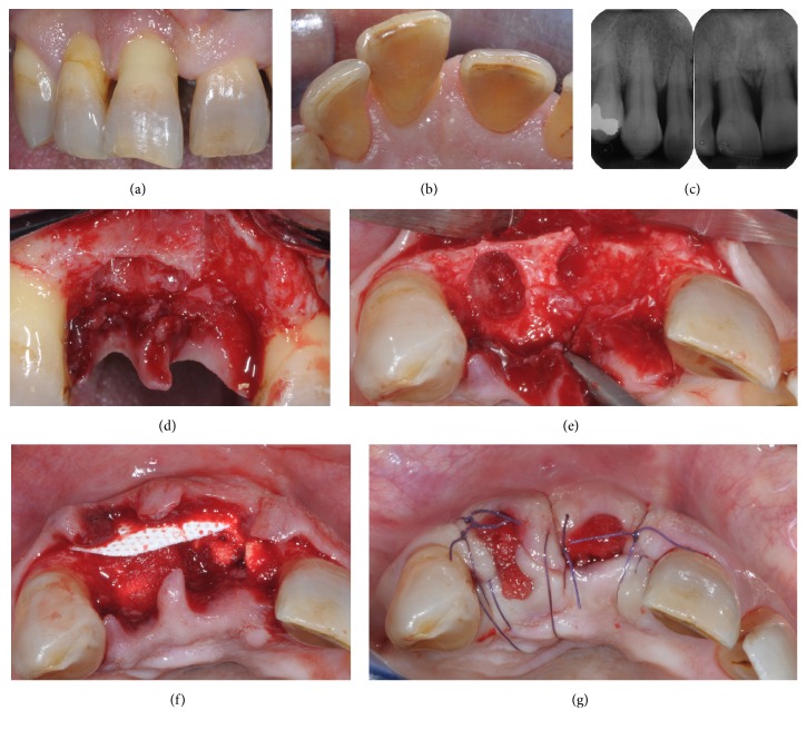
Figure 1.
Maxillary right central and lateral incisors before extraction (a). Note extrusion of central and lateral incisors, migration of central incisor, and persistent inflammation (b). Preoperative radiographs showing severe interdental bone loss and widening of the residual periodontal ligament space as a consequence of the occlusal trauma (c). Intraoperative view following teeth removal (d). Occlusal view showing vertical bone resorption and partial nonspace maintaining defect at central incisor (e). After placement of a nonresorbable d-PTFE membrane to replace the missing buccal bony wall, the gap between the membrane and the residual palatal wall was filled with the collagen sponge (f). Flaps were repositioned and the membrane was left partially exposed and protected with the collagen sponge (g).

