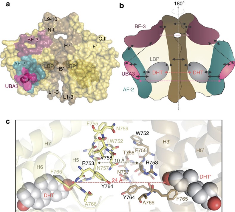
Figure 7. Proposed allosteric communication pathways across the AR-LBD dimer interface.
(a) Surface representation of the AR homodimer. The dimer interface (brown), the AF-2 groove (blue) and the BF-3 pocket (raspberry) are highlighted. Residues that form or line the LBP are shown with a Connolly dot surface and the UBA3 peptide as a pink surface. (b) Schematic representation of the proposed intra- and inter-domain allosteric pathways in AR-LBD. Solid arrows indicate short-range communication networks, while dashed arrows point to long-range interactions. (c) Close-up of the dimer interface highlighting allosteric communication between the LBPs across the dimer interface. The distances between the two R753 residues and the two DHT moieties are given.

