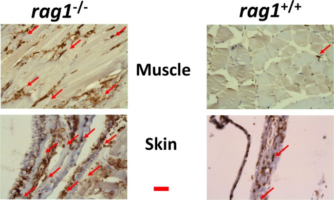
Figure 5.

Leukocyte infiltration in muscle and skin tissues in rag1−/− and rag1+/+ adult zebra fish. Histological sections from muscle and skin tissues of rag1−/− and rag1+/+ were stained with an antibody anti-l-plastin, a pan-leukocyte marker (dark-stained areas). Red arrows, examples of l-plastin stained cells. Images are representative of at least two independent experiments. Horizontal red line, 30 μm.
