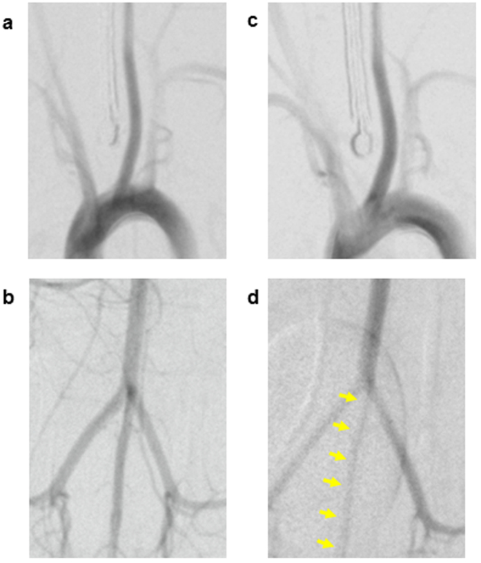
Figure 4. In vivo digital subtraction angiography showing a spastic caudal ventral artery after tail cooling in rats.
(a,b) The left common carotid artery (a) and the caudal ventral artery (b) were visualised in the control (HwTw) group. (c) The left common carotid artery was not spastic in the head cooling (HcTw) group. (d) The caudal ventral artery was spastic (arrows) in the tail cooling (HwTc) group. Thus, tail cooling in rats can easily induce angiospasm of the tail artery.

