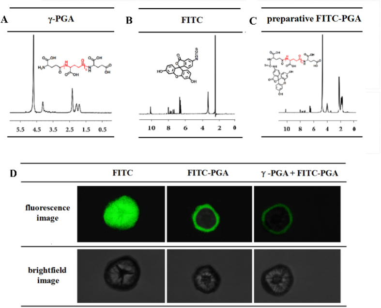
Figure 2. The fluorescence tracing of poly-γ-glutamic acid (γ-PGA) in Brassica napus root protoplasts.
(A) The 1H-NMR spectra of γ-PGA; (B) the 1H-NMR spectra of fluorescein isothiocynate (FITC); (C) the 1H-NMR spectra of preparative FITC-PGA; (D) laser scanning confocal microscope (LSCM) scanning images of root protoplasts after treatment with FITC, FITC-PGA, or γ-PGA plus FITC-PGA, respectively.

