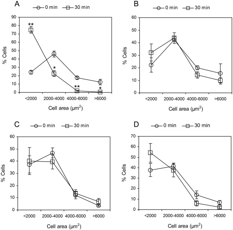
Figure 5. Cell area distributions of HUVSMCs with or without treatment exposed to static or 30-min 20 dyn/cm2 shear stress condition.
(A) Control cells without treatment exposed to shear. (B) Control cells without treatment exposed to static. (C) HUVSMCs pre-treated with 0.2 U/ml Hep.III for 1 hour exposed to shear. (D) HUVSMCs pre-treated with 10 μM Y-27632 for 1 hour exposed to shear. *P < 0.05 vs. time 0. **P < 0.01 vs. time 0.

