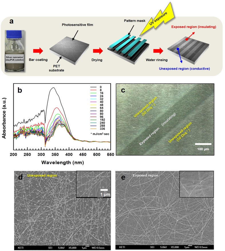
Figure 1.
(a) The preparation procedure for the patterned transparent electrode layer with a continuous distribution of AgNWs. (b) The dependence of the UV absorbance on the exposure dose. (c) The confocal microscopy image of the patterned layer. (d,e) The SEM images of the unexposed and exposed regions in the patterned layer, respectively. The inset of each image is a magnification.

