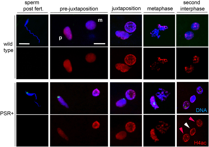
Figure 2. Acetylated histone H4 patterns are normal in PSR-carrying embryos.
In wild type embryos, histone H4 acetylated at multiple Lysine residues is absent from the sperm’s chromatin immediately after fertilization. However, H4ac appears on the paternal chromatin as the sperm and egg nuclei migrate toward one another (pre-juxtaposition); the maternal nucleus and meiotic products (not shown) already show H4ac. This signal increases slightly more on the paternal set compared to the female set during metaphase, but by the end of the first mitosis both daughter nuclei contain similar levels of H4ac. In PSR-carrying embryos the patterns are indistinguishable. The paternal chromatin mass (PCM, indicated by white arrowhead in the bottom right panel) contains the same level of H4ac as the maternally derived nuclei (red arrowheads). PSR is highlighted by a sequence-specific FISH probe (green in panels of the bottom two rows). Scale bar equals 5 μM in the top left panel and 12 μM in the adjacent panel to the right, under pre-juxtaposition. p and m stand for paternal and maternal nuclei.

