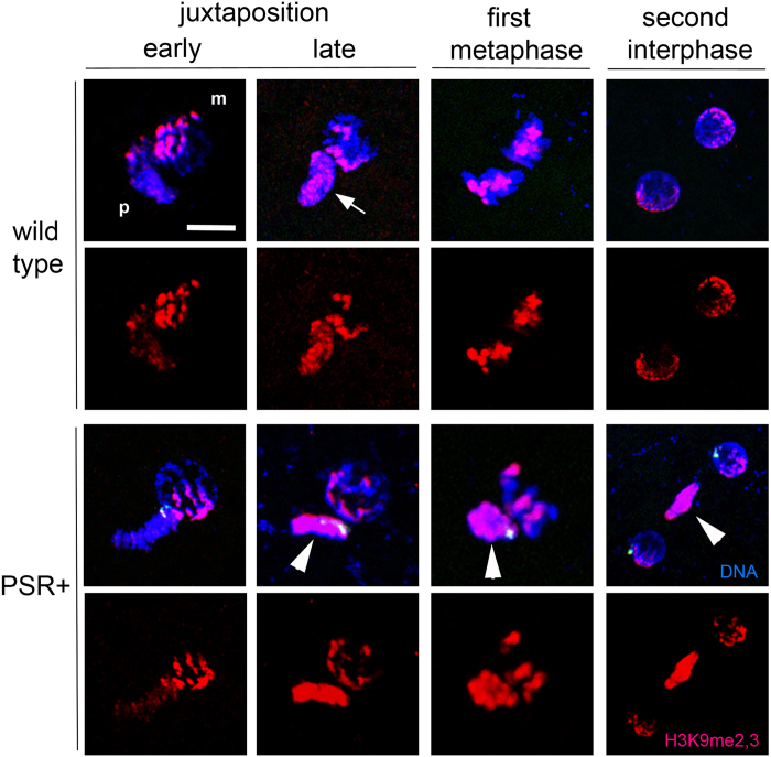
Figure 3. PSR disrupts histone H3K9me2 and H3K9me3 patterns on paternally-derived chromatin.
In wild type embryos H3K9me2,3 begins to appear in localized regions of the paternal chromatin following juxtaposition. The signal persists in these regions while increasing in intensity (white arrow) until they are similar to regions on the maternal chromatin. This mark persists during metaphase, and obtains a RABL-like confirmation in the pericentromeric regions on opposing sides of the two daughter nuclei at the end of the first mitosis. In PSR-carrying embryos, the paternal chromatin obtains abnormally high levels of H3K9me2,3 across its entirety instead of in distinct regions (white arrowheads). This pattern persists following the end of the first mitosis. Scale bar is 10 μM.

