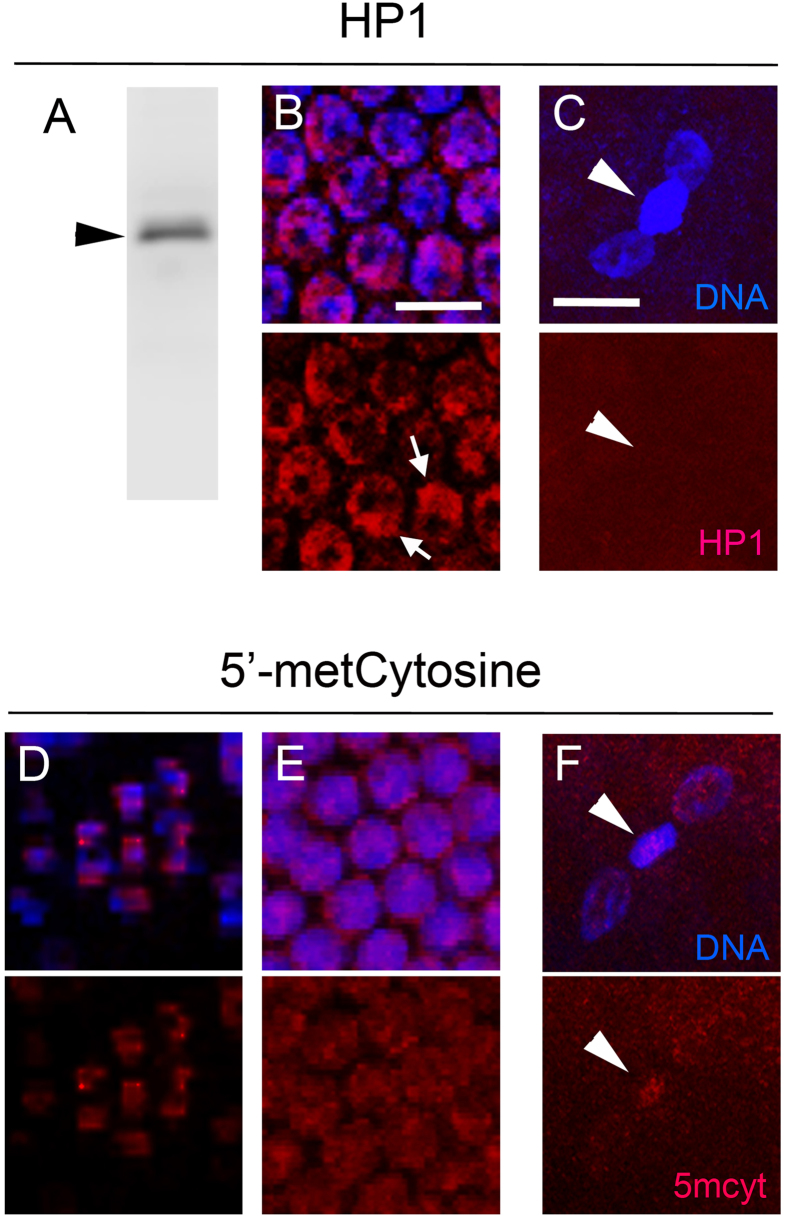
Figure 4. PSR does not cause ectopic heterochromatinization of the paternal chromatin or grossly affect its 5′-methylCytosine pattern.
(A) A polyclonal serum raised against the single N. vitripennis HP1 protein recognizes a single band of 24 kDa (black arrowhead), its predicted mobility size, by Western blot. (B) The serum highlights bright, chromocenter-like regions (white arrows) within the nuclei of embryos in late cleavage. (C) However, no HP1 is present on either the PCM (white arrowheads) or maternally derived daughter nuclei following the first mitotic division. A monoclonal antibody highlights low but visibly detectable levels of 5′-methylCytosine in the nuclei of spermatogonia (D) and late cleavage embryos (E). (F) 5′-methylCytosine levels were slightly enriched on the PCM (white arrowhead) compared to the maternally derived nuclei at the end of the first mitotic division. Scale bars in (B) and (C) equal 10 μM and 12 μM, respectively.

