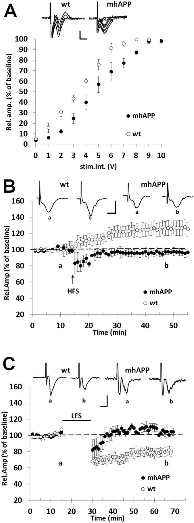
Figure 2. Synaptic plasticity impairment in EC slices from 6 month old mhAPP mice.

A significant difference was observed in basic synaptic transmission between WT and mhAPP (A) the plot represents the relationship between the amplitude of the response and the stimulus intensity under basal conditions (input–output curve). In (C) long term potentiation (LTP) was normally expressed in EC slices from WT mice (open circles). In contrast, LTP magnitude was not inducible by HFS in mhAPP slices (black circles); insert shows representative FPs recorded during baseline (a) or after HFS stimulation (b). Moreover, LFS stimulation (D) was not capable of modifying FPs amplitude in EC slices from 6 month old mhAPP mice (black circles), whereas it was capable of inducing the LTD in WT slices (open circles); insert shows representative FPs recorded during baseline (a) or after LFS stimulation (b). In (C,D) scale bars correspond to 0.5 mV and 5 ms.
