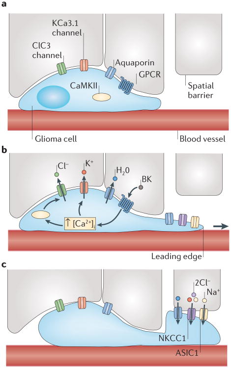
Figure 2. A hydrodynamic model of glioma cell migration.
a | Glioma cells encounter spatial barriers (for example, other cells) as they migrate along blood vessels through the brain. Gliomas express various channels, including Cl− channel protein 3 (ClC3; a voltage-gated Cl− channel), Ca2+-activated K+ channel KCa3.1 and aquaporins. b | Upon stimulation of G protein-coupled receptors (GPCRs), there is a rise in intracellular Ca2+ concentration ([Ca2+]i) in glioma cells, leading to Ca2+-dependent activation of Ca2+/calmodulin-dependent protein kinase II (CaMKII)-dependent ClC3 and KCa3.1 channel opening. This leads to the efflux of Cl− and K+, obligating water to flow down its osmotic gradient and leave the cell. This volume decrease enables glioma cells to decrease cytoplasmic volume and squeeze into small spaces. c | After passing a spatial barrier, glioma cells can regain volume through ion influx through the Na+–K+–Cl− cotransporter 1 (NKCC1) and acid-sensing ion channel 1 (ASIC1). BK, bradykinin.

