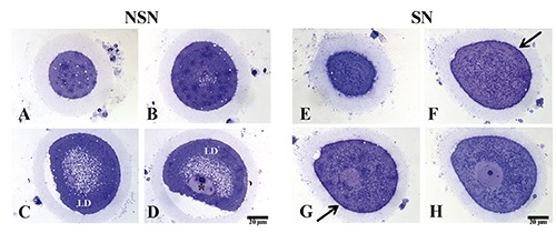
Figure 2.

A-D) Representative images of semi-thin sections of discarded human NSN oocyte; asterisk refers to heterochromatin blocks; LD, lipid droplets. E-H) Representative images of semi-thin sections of discarded human SN oocyte; arrows point to thin glycocalix distributed at the cellular surface.
