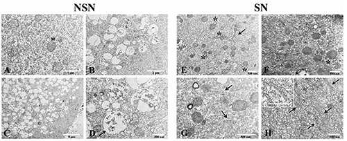
Figure 3.

A-D) Transmission electron microscopy images of discarded human NSN oocyte; asterisk refers to mitochondria with microvacuolized matrix and both external and internal membrane protrusion; arrow points to residual ghosts of mitochondrial lysis. E-H) Transmission electron microscopy images of discarded human SN oocyte. Asterisks refer to mitochondrial vacuolar protrusions; arrow in E points to Golgi apparatus and arrows in G refer to smooth/rough reticulum; cytoplasmic lattices are visible in H (arrows); insert in H shows a magnification of CPLs.
