Fig. 6.

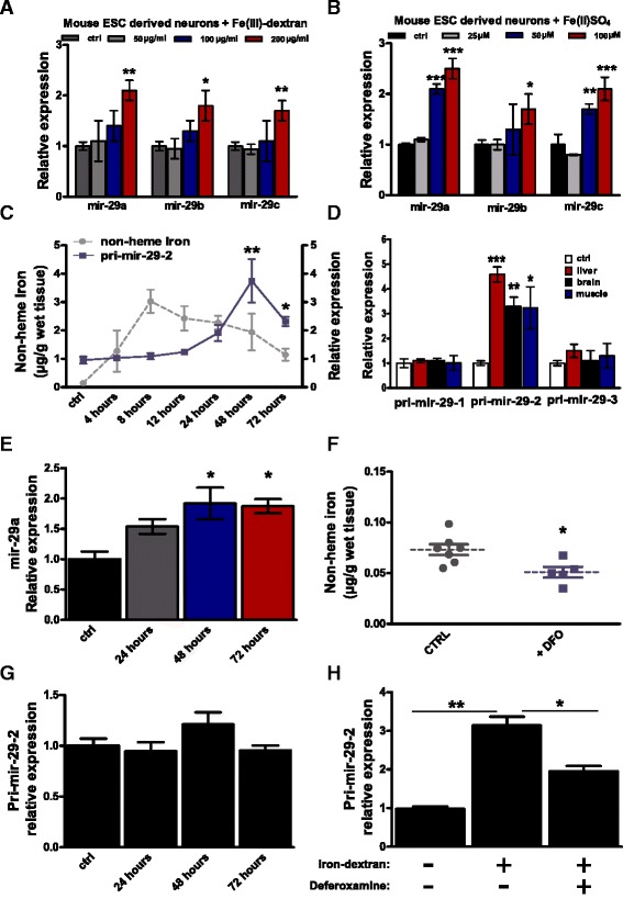
Iron overload induces miR-29 up-regulation in neurons and brain. a, b Modulation of miR-29 family members in murine neurons derived from mESCs incubated respectively with a 0, 50, 100, or 200 μg/mL of Fe(III)-dextran or with b 0, 25, 50, or 100 μM of Fe(II)SO4 for 72 hours (* P < 0.05; **P < 0.01; ***P < 0.001, one-way ANOVA with post-hoc Tukey’s test), n = 3 independent experiments performed in duplicates for each condition. c Time course of non-heme iron amount (quantified by colorimetric analysis) and pri-miR-29-2 expression level in fish brain (quantified by RT-qPCR) following intraperitoneal (i.p.) injection of 350 μg/g of iron dextran, control animals were injected with saline solution. Grey line represents iron amount, blue line represents pri-miR-29-2 relative expression level (setting the baseline to 1, *P < 0.05; **P < 0.01; one-way ANOVA with post-hoc Tukey’s test), n = 4 biological replicates for each point. d Relative expression of miR-29 primary transcripts (pri-mir-29-1, 2, 3) in liver, brain and muscle 48 hours following i.p. injection of 350 μg/g of iron dextran. Data were normalized on TBP expression and control animals relative expression was considered as baseline (***P < 0.001; **P < 0.01; *P < 0.05; Mann–Whitney U-test), n = 5 biological replicates. e Expression of mature N.fu-miR-29a at 48 hours after iron injection quantified by RT-qPCR (* P < 0.05; Mann–Whitney U-test), n = 4 biological replicates for each time point, U6 was used as normalization control. f Non-heme iron quantification 24 hours after deferoxamine (DFO) i.p. injection, control animals were injected with saline solution (*P < 0.05; Mann–Whitney U-test), n = 7 control animals and n = 5 DFO injected animals. g Time course of pri-miR-29-2 expression level (quantified by RT-qPCR) following i.p. injection of 30 μg/g of DFO, one-way ANOVA P = 0.1663, n = 4 biological replicates for each time point. h Modulation of pri-miR-29-2 expression in fish brain (quantified by RT-qPCR) after iron overload and in combination with administration of 30 μg/g of DFO, control animals were injected with saline solution (*P < 0.05; Mann–Whitney U-test), n = 5 biological replicates. For all the graphs, mean ± standard errors of means are reported
