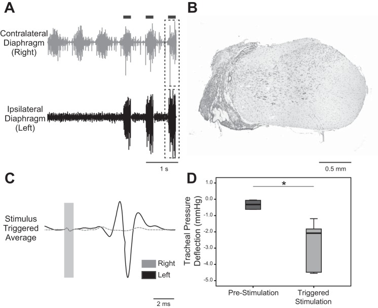
Fig. 5.
ISMS after subacute C2Hx. A: representative diaphragm EMG activity after subacute C2Hx. In the example traces, genioglossus-triggered ISMS was delivered during the breaths marked by the black bars. B: histological section of the C2 spinal cord stained with cresyl violet. The example demonstrates an anatomically complete hemilesion extending to the midline of cervical cord. C: stimulus-triggered averages from the ipsilesional (solid line) and contralesional diaphragm (dashed line); data were obtained from the period indicated by the dashed boxes in A. These traces illustrate activation of the diaphragm ipsilateral to the C2Hx lesion. D: average change in tracheal pressure during lung inflation at prestimulation baseline and during ISMS. *P < 0.05.

