Fig. 1.
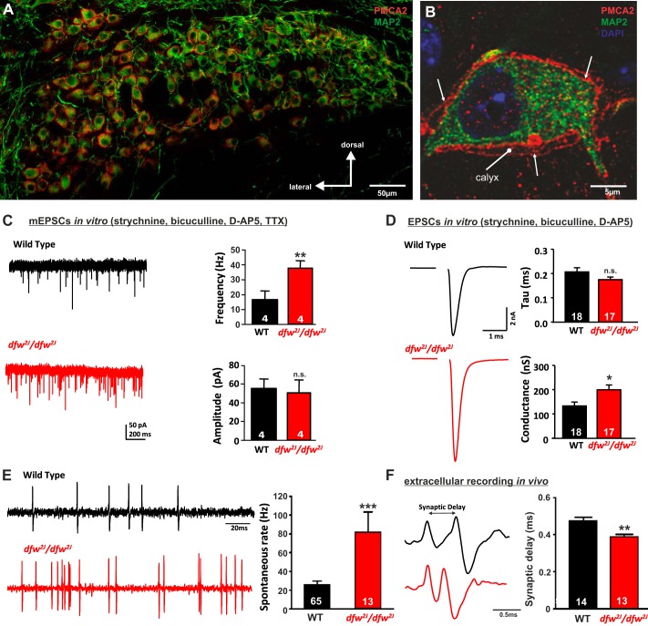
PMCA2 regulates transmitter release in the MNTB. A and B: immunohistochemical labeling for MAP2 and PMCA2 in the MNTB (A) and in an individual MNTB neuron (B). A cross section through the calyx is marked “calyx.” Arrows show where PMCA2 appears to be localized presynaptically in the outer membrane of the calyx. C: voltage-clamp recordings from postsynaptic MNTB neurons in acute brain slices show a higher frequency of miniature excitatory postsynaptic currents (mEPSCs) in the MNTB of dfw2J/dfw2J mice compared with wild type (WT). D: calyceal EPSCs evoked by midline stimulation are larger in dfw2J/dfw2J mice compared with WT. Stimulus artifacts have been deleted for clarity. WT data include 7 medial cells, 3 lateral cells, and 8 cells with no information about location in the MNTB. dfw2J/dfw2J data include 7 medial cells, 6 lateral cells, and 4 cells with no information about location in the MNTB (see also Fig. 4C). E and F: in vivo single-unit recordings of MNTB neurons measured higher spontaneous firing rates (E) and shorter synaptic delays (F) in dfw2J/dfw2J mice compared with WT. ***P ≤ 0.001, **P ≤ 0.01, *P ≤ 0.05, n.s., Not significant.

