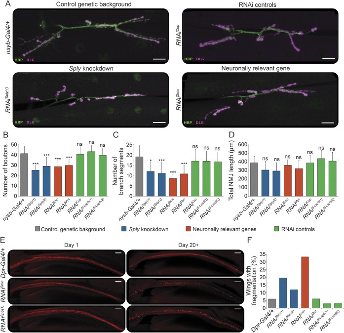
Figure 3. Analysis of neuron-specific Sply deficiency in Drosophila.
(A) Immunolabeling of presynaptic (anti-HRP antibody, green) and postsynaptic (anti-DLG antibody, magenta) compartments of the neuromuscular junctions (NMJs) in third instar larvae, pan-neuronally (nsyb-Gal4) expressing RNAi constructs against Sply (Sply knockdown; RNAiSply[1], RNAiSply[2]), neuronally relevant genes (RNAiSmn, RNAiMyc), genes not expressed in neurons (RNAi controls; RNAiCup, RNAiS-Lap5[1], RNAiS-Lap[2]), or the driver alone (control genetic background; nsyb-Gal4/+). Scale bar, 20 μm. (B–D) Quantitative analysis of the NMJ phenotypes. Maximum intensity projections of Z-stacks comprising the full NMJ were used. The number of boutons per NMJ (B) was counted using the Cell Counter plugin. ImageJ was used to count the number of individual branch segments (C) and calculate the total NMJ length (D). Error bars represent the SD of at least 13 NMJs per genotype. *p < 0.05; ***p < 0.001; ns = nonsignificant (one-way analysis of variance with Bonferroni multiple comparison test against the control genetic background). (E) Nerve tract along the wing L1 vein visualized by mCherry, where RNAi constructs are expressed using the dpr-Gal4 driver. Representative images of wings are shown for day 1 and day 20+ old flies expressing the driver alone (Dpr-Gal4/+), neuronally relevant gene (RNAiSmn), or Sply (RNAiSply[1]). Scale bar, 20 μm. (F) Quantification of the number of wings showing axonal fragmentation per genotype (n = 30–60/genotype).

