
Fig. 1.
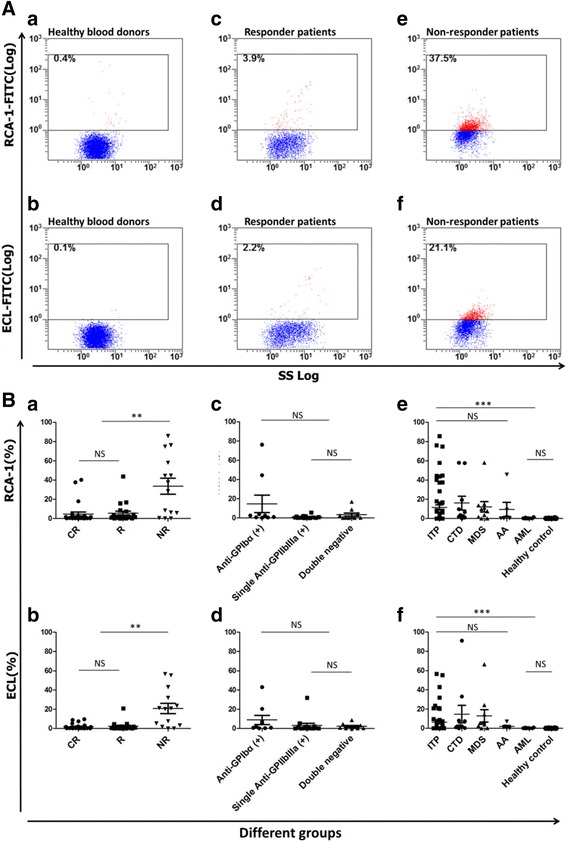
A Platelet desialylation in representative patients and healthy controls. The RCA-1 and ECL binding to platelets from healthy blood donors (a, b), responder (c, d), and non-responder patients (e, f) were examined by flow cytometry. The representative dot plots from each group are shown. B Platelet desialylation in different patient groups and healthy controls. The RCA-1 and ECL levels (Mean ± SEM) in complete responders (CR), responders (R) and non-responders (NR) (a, b); anti-GPIbα antibody positive (+) group, single anti-GPIIb/IIIa antibody (+) group, and double negative group (c, d); and in the thrombocytopenias/controls (e, f) (ITP, CTD, MDS, AA, AML, and healthy controls) were examined by flow cytometry. Each point represents the level of platelet desialylation of an individual patient or healthy blood donor (**p < 0.01; ***p < 0.001)